Foot overview
Radiographs
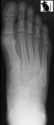
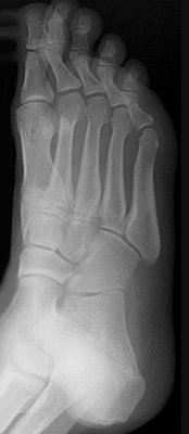
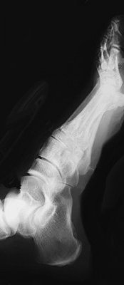
Standard views - AP, Oblique, Lateral
 |
 |
 |
|---|
(Mouse over Ap view)
To exclude midfoot/ lisfranc injuries make sure
- The 2nd metatarsal lines up with the medial border of the middle cuneiform on the AP view
- The 4th metatarsal lines up with the medial border of the cuboid on the oblique
- Consider weight bearing films
Look for:
- Base of 5th metatarsal fracture
There are many accessory bones in the foot that may be mistaken for fractures. Correlate areas of tenderness to abnormal areas.
Special circumstances
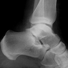
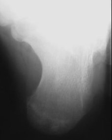
Calcaneus
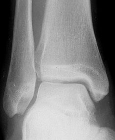
Views needed AP and lateral ankle and axial view
 |
 |
 |
| AP Ankle | Lat Ankle | Axial View |
Look for/ at:
- Exclude ankle fracture
- Decide if intra articular (subtalar joint, calcaneocuboid joint or extraarticular)
- Bohlers angle
- Crucial angle Gisane
- On Axial view look at shortening and widening of heel
See essex lopressti for evaluation of calcaneus
Talus
- Need AP and lateral ankle to see fracture and ensure talus congruent in mortise
- Modified AP of foot -Canale and Kelly described technique to demonstrate the entire talar neck in the anterior-posterior direction. Ankle in max equinus, place foot on cassette and pronate 15 degrees, direct x-ray tube cephalad at 75-degree angle from the horizontal.