Calcaneus fracture
Usually follows fall from height. Beware of further injuries to axial skeleton (T and L spine 10%). Bilateral in 5-9%. Counsel patient regarding severity of injury. Treatment is still controversial especially for intra articular fractures, surgery does not always improve function and risks wound complications (older 60 and smoking)
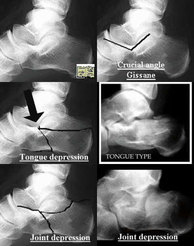
Classification
Essex Lopressti (1952)
 |
|
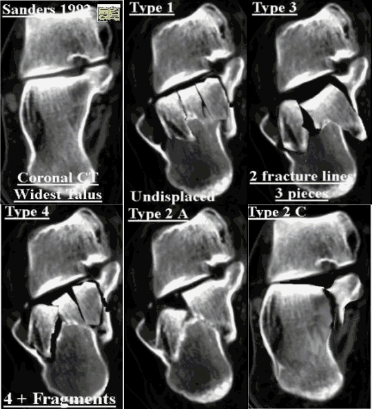
Sanders CT Classification
 |
The section showing the widest undersurface of the posterior facet is arbitrarily used. The talus isdivided into three equal columns by two lines.A third line is drawn just medial to the medial edge of the posterior facet. Dividing the posterior facet into three potential pieces: a medial, central, lateral and a sustentacular fragment. |
Radiology
Ap and Lateral calcaneum (essentially AP foot and Lateral ankle centred on Calcaneum)
Axial view calcaneum
AP ankle (useful to exclude malleolar fracture and show degree of peroneal impingement laterally)
Special views
| LatOblique | Medial border of foot placed on, sole inclined 45°. X-ray centered 2.5 cm below and 2.5 cm ant. to lat. malleolus. Allows visualization of ant. facet of the subtalar joint and ant. calcaneal process |
| Med. Oblique Axial | Foot dorsiflexed and inverted,
30° foam wedge on the x-ray, limb internally rotated
60°. X-ray centered 2.5 cm below and 2.5 cm anterior to
tip of lat. malleolus and tilted 10° cephalad.
Demonstrates both the middle and post. facets. Brodén simillar internally rotated 45°. X-ray tube angled 40°, 30°, 20°, and 10° cephalad |
| Brodén view | foot in neutral flexion, leg internally rotated 30 to 40 degrees. X-ray centered over lat. malleolus and four radiographs are made, with the tube angled 40, 30, 20, and 10 degrees toward the head of the patient. |
| Lat. Oblique Axial | Foot dorsiflexed and everted limb externally rotated 60°, lateral border lies against 30° foam wedge.X-ray tube centered 2.5 cm below the med. malleolus, tilted 10° cephalad. Shows posterior facet |
Computerised Tomography
CT is important in evaluating calcaneal fractures. It is invaluable if surgery is contemplated. Several classification systems have been described.
Management
Varies if intra articular or extra articular. Extra articular generally non operative except for "parrot beak" avulsion fracture
If heel widened consider compressing to avoid peroneal impingement. Intra articular treatment controversial still. Options (1) no reduction and early motion; (2) closed reduction and fixation; (3) open reduction and grafting or internal fixation; and (4) primary arthrodesis.
Problems with fracture:
- Heel widens and shortens, patients find it hard to fit shoes
- Widening of calcaneum leads to lateral impingement on the peronei below the lateral malleolus
- Involvement of subtalar joint, and calcaneocuboid joint leads to subtalar stiffness and pain.
Admit for elevation and analgesia these hurt and swell up (10% compartment syndrome). Exclude associated injuries actively, if any back pain image it.
If considering surgery arrange CT.
Remain non weight bearing 6-12 weeks till union. Encourage patients improvement may continue for up to 18 months
Current Concepts Review - Displaced Intra-Articular Fractures of the Calcaneus -The Journal of Bone and Joint Surgery (A) 82:225-50 (2000)
Operative Compared with Nonoperative Treatment of Displaced Intra-Articular calcaneal fractures - The Journal of Bone and Joint Surgery (American) 84:1733-1744 (2002)
Summary - "prospective, randomized, controlled multicenter trial demonstrated that operative treatment as a whole provides no improvement over nonoperative treatment of displaced intra-articular calcaneal fractures. However, careful stratification of the patient population and clinical outcome information distinguishes certain features that support surgical care for displaced intra-articular calcaneal fractures. Statistical analysis demonstrated that women, patients who were not receiving Workers' Compensation, younger males, patients with a higher Böhler angle, patients with a lighter workload, and those with a single, simple displaced intra-articular calcaneal fracture have better results after operative treatment than after nonoperative treatment. Anatomic or near anatomic reductions enhance outcomes while comminuted reductions or fractures without reduction produce long-term outcomes that are less satisfactory. Nonoperative care more commonly leads to late arthrodesis. The best patients to treat nonoperatively are those who are fifty years old or more, males, and those who are receiving Workers' Compensation and have an occupation involving a heavy workload. The results after a higher-energy fracture (a lower Böhler angle and more comminution) are not as good as those after a low-energy injury".