Extended anterolateral - Shoulder
Indication
-
Open reduction and internal fixation proximal humeral fractures
-
Hemiarthroplasty for trauma (usually don't need to extend below axillary nerve see antero superior, mckenzie)
The deltopectoral approach to the shoulder requires extensive soft tissue dissection and muscle retraction to gain adequate exposure of the lateral aspect of the humerus.
Extensive dissection anteriorly may
further compromise humeral head blood supply (See,
anterior circumflex humeral
artery, humeral head blood supply).
With any deltoid splitting approach, there are concerns regarding injury to the
axillary nerve with subsequent
dysfunction of the anterior deltoid.
Historically the distal split has been restricted to 3 – 6cm distal to the acromion.
Mackenzie described splitting the deltoid no further than 6 cm distally from the lateral edge of the acromion then inserting a stay-suture to avoid potential injury to the nerve.
Cadaveric work has clearly deliniated the path of the axillary nerve in relation to the acromion and and prominence of the greater tuberosity.
The axillary nerve is usually easily palpable as it exits the quadrilateral space and travels approximately 35 mm from the prominence of the greater tuberosity in the line of the deltoid raphe.
Anatomy
The axillary nerve arises from the posterior cord of the brachial plexus and passes through the quadrilateral space, dividing into anterior and posterior branches.
The anterior motor branch wraps around the neck of the humerus and gives variable innervation to the three heads of the deltoid.
At the junction of the anterior and middle heads of the deltoid exists the avascular anterior raphe (vascular watershed).
The anterior motor branch of the axillary nerve crosses the humerus transversely
at variable distances as a single nerve and penetrates the fascia of the deltoid
before or after dividing into several smaller branches.
| Author | n | Acromion | Greater tuberosity |
| Duparc et al |
32
|
Undersurface acromion, anterior branch, after dividing the raphe - 63.3 mm | 35.5 mm distal to the lateral prominence of the greater tuberosity |
| Burkhead |
102
|
Anterolateral acromion, average 57 cm (range, 41–71 mm) | |
| Vathana et al | 77 | 63 mm from the acromion | |
| Lin et al | 20 | Prominence of the greater tuberosity, (average 45.6 mm; range, 34–54 mm) | |
| Gardner et al | 20 | Undersurface acromion - superior border anterior branch at raphe, ave. 63.3 mm (range, 53.2–70.4 mm; SD 4.9 mm; 99% confidence interval, 60.2–66.4 mm) | Prominence greater tuberosity, ave. 35.5 mm (range, 32.1–42.5 mm; SD 2.3 mm; 99% confidence interval 34.0–37.0) |
Vathana et al pointed out the
acromion is not parallel to axillary nerve and as such suggested it is not a
good surgical landmark for the nerve.
The standard deviation is less when
measuring from the greater tuberosity than from the acromion(2.3 versus 4.9mm )
However when using the extended anterolateral acromial approach, the nerve is identified in the line of the raphe, and in fracture surgery the greater tuberosity anatomy is distorted.
As such the acromion may be an adequate landmark.
Gardner et al showed the first branches of the anterior branch arise at least 4 mm posteriorly and 5 mm anteriorly to the raphe, so that using an incision that does not stray from the raphe will not place other branches at risk.
The superior obliquity of the path of the nerve is approximately 20°, and the width of the nerve as the nerve crosses the raphe is 4.2mm.
The posterior humeral circumflex artery passes through the quadrilateral space with the axillary nerve.
Considerations
If performing a hemiarthroplasty for trauma you normally don't need to extend below the axillary nerve, but have to release the anterior deltoid of the acromion to get access.
Positioning
Beach-chair (see shoulder arthroscopy positioning)
Skin Incision
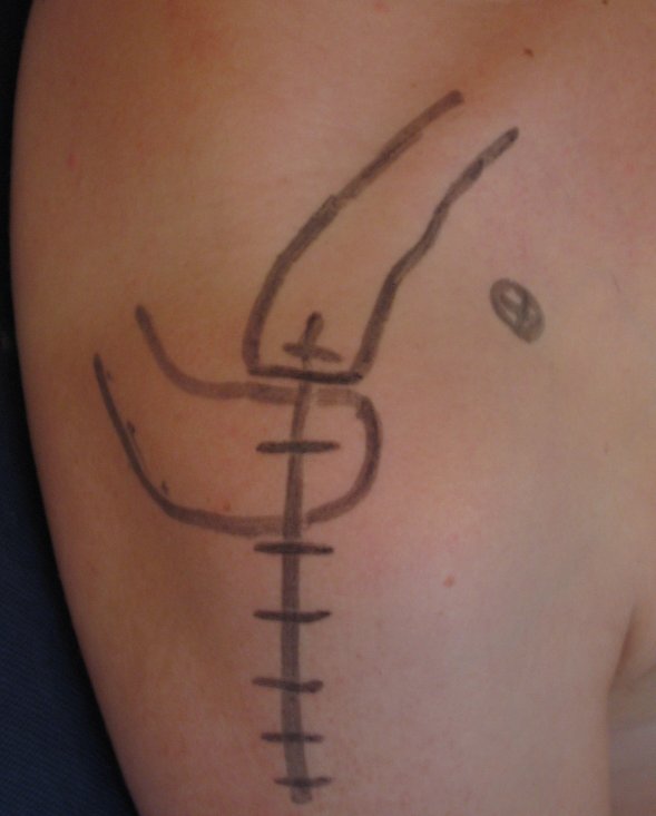 |
Identify/ mark out: spine of scapula, acromion, acromioclavicular joint, lateral clavicle and coracoid. Longitudinal incision, starting over the anterior acromion running distally down the deltoid for 5 - 7 cm (skin deep) from the lateral edge of the acromion. |
Superficial dissection
Elevate skin flaps as needed to identify the raphe between the anterior and middle deltoid and split the raphe in the line of the fibres distally for 2 - 4 cm from acromion.
Insert a finger into the subacromial and deltoid bursa, flex and sweep it around to locate the cord like structure of the axillary nerve on the under surface of the deltoid. (Flatow, Duval).
Once the axillary nerve has been palpated to obtain a general idea of its
location, extend the incision in the raphe carefully till the nerve and
posterior humeral circumflex artery are identified and isolated.
You can now develop 2 windows, one above and one below the axillary nerve.
To improve proximal access elevate 1- 2 cm of anterior deltoid off anterior acromion, subperiostealy.
Consider performing an anterior acromioplasty.
Deep dissection
Identify and mobilize the fracture fragments:
-
Greater tuberosity, posterosuperior, under the acromion in line with the pull of the
supraspinatus/infraspinatus, teres minor. -
Lesser tuberosity, anteromedial, pull of subscapularis.
If needed identify and split the rotator interval in line with the cuff fibres.
In 3 and 4 part fractures, the head is typically lying medially and facing superiorly.
Place stay sutures through tendinous insertion on tuberosities
Elevate and reduce the head to its correct position.
Reduce and suture the tuberosities behind the head.
Once all fragments are reduced and secured, apply a plate to the lateral side of the humeral head/shaft.
When applying a plate, the axillary nerve is mobilized and elevated, allowing the plate to be slipped underneath the nerve.
Fracture fixation with a plate and screws can then proceed as normal.
Exposure extension
To increase proximal exposure the anterior deltoid can be elevated subperiostealy of the anterior acromion.
Closure
If the anterior deltoid has been released from the acromion a secure repair is needed.
Consider trans-osseous sutures through the acromion using a 1 PDS on a short J needle.
Close the deltoid raphe
Post operatively
Rehab as for internal fixation of proximal humeral fracture.
References
Gardner, Michael J MD; Griffith, Matthew H MD; Dines, Joshua S MD; Briggs, Stephen M PA; Weiland, Andrew J MD; Lorich, Dean G MD The Extended Anterolateral Acromial Approach Allows Minimally Invasive Access to the Proximal Humerus. Clinical Orthopaedics & Related Research. 434:123-129, May 2005.
Techniques in Shoulder and Elbow Surgery 7(2):77–81, 2006; An Anterosuperior Approach for Proximal Humeral Fractures; Mark Webb, FRCS (T&O) and Lennard Funk.
Mackenzie D. The anterior-superior
approach to the shoulder.
Orthop Trauma. 1993;2:71- 77.
Flatow EL, Bigliani LU. Tips of the trade. Locating and protecting the axillary nerve in shoulder surgery: the tug test. Orthop Rev. April 1992;21(4):503Y505.
Duval MJ, Parker AW, Drez Jr D, Hinton MA: The anterior humeral circumflex vessels and the axillary nerve: An anatomic study. Orthop Rev 22:1023–1026, 1993.