Antero superior - shoulder
(Mckenzie)
Indication
-
Total shoulder replacement
-
Shoulder resurfacing arthroplasty
-
Hemiarthroplasty for trauma
-
With extended anterolateral approach open reduction and internal fixation of proximal humeral fractures
Provides access to:
-
Gleno-humeral joint
-
Humeral head and tuberosities
-
Acromion
-
AC joint
Anatomy
The axillary nerve is at risk at the inferior extent of the deltoid split.
The axillary nerve arises from the posterior cord of the brachial plexus and passes through the quadrilateral space, dividing into anterior and posterior branches.
The anterior motor branch wraps around the neck of the humerus and gives variable innervation to the three heads of the deltoid.
At the junction of the anterior and middle heads of the deltoid exists the avascular anterior raphe (vascular watershed).
The anterior motor branch of the axillary nerve crosses the humerus transversely
at variable distances as a single nerve and penetrates the fascia of the deltoid
before or after dividing into several smaller branches.
| Author | n | Acromion | Greater tuberosity |
| Duparc et al |
32
|
Undersurface acromion, anterior branch, after dividing the raphe - 63.3 mm | 35.5 mm distal to the lateral prominence of the greater tuberosity |
| Burkhead |
102
|
Anterolateral acromion, average 57 cm (range, 41–71 mm) | |
| Vathana et al | 77 | 63 mm from the acromion | |
| Lin et al | 20 | Prominence of the greater tuberosity, (average 45.6 mm; range, 34–54 mm) | |
| Gardner et al | 20 | Undersurface acromion - superior border anterior branch at raphe, ave. 63.3 mm (range, 53.2–70.4 mm; SD 4.9 mm; 99% confidence interval, 60.2–66.4 mm) | Prominence greater tuberosity, ave. 35.5 mm (range, 32.1–42.5 mm; SD 2.3 mm; 99% confidence interval 34.0–37.0) |
Vathana et al pointed out the
acromion is not parallel to axillary nerve and as such suggested it is not a
good surgical landmark for the nerve.
The standard deviation is less when
measuring from the greater tuberosity than from the acromion(2.3 versus 4.9mm )
However in fracture surgery the greater tuberosity anatomy is distorted.
As such the acromion may be an adequate landmark.
The posterior humeral circumflex artery passes through the quadrilateral space with the axillary nerve.
Considerations
Positioning
Beach-chair (see shoulder arthroscopy positioning)
Shoulder arthroplasty as per constant
Skin Incision
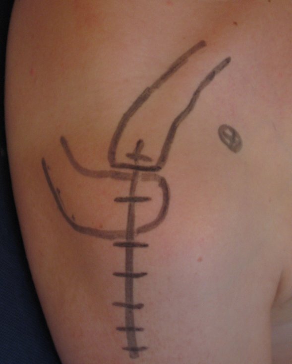 |
The skin incision extends distally in a straight line from just posterior to the acromioclavicular joint for a distance of 9 cm. |
Superficial dissection
The anterior deltoid fibers are split for a distance of not more than 6 cm.
Place a 1 vicryl stay suture in the distal end of the split to prevent further extension and possible injury to the axillary nerve.
The acromial attachment of the
deltoid is lifted with an osteo-periosteal flap to expose the anterior acromion
and preservation of the superior acromioclavicular ligament.
An anterior acromioplasty according to the technique of Neer is performed.
Deep dissection
If performing an artroplasty:
The shoulder is flexed and externally rotated to facilitate coagulation of the anterior circumflex humeral vessels.
The rotator interval is identified and longitudinally incised along the line of the long head of biceps to identify the exact insertion of subscapularis.
Stay sutures are placed into the subscapularis.
The tendon is divided 2 cm medial to the bicipital groove.
If the subscapularis appears tight
consider dividing it in an oblique or “Z” manner to allow repair with
lengthening of the tendon.
Release the joint capsule anteriorly and inferiorly whilst taking care to
protect the axillary nerve with a blunt
elevator where it passes through the quadrilateral space.
The glenohumeral joint may now be dislocated anteriorly by external rotation and
extension, allowing full exposure of the humeral head and neck.
Perform arthroplasty/ procedure.
Exposure extension
If further exposure is needed, then excision of the lateral end of 1cm of clavicle considerably enhances exposure.
For distal extension below axillary nerve in trauma cases (ORIF) see extended anterolateral approach.
Closure
The subscapularis is repaired using No.1 suture material (absorbable (PDS) or
non-non absorbable) without plicating the subscapularis or with through bone
sutures.
The rotator interval is closed.
If there is any rotator cuff
deficiency and this is repairable then full rotator cuff repair is made in the
normal manner at this stage. Every attempt is made to close the rotator cuff
completely.
The deltoid is reattached to the acromion with No. 1 absorbable sutures (PDS J
needle) through bone.
The deltoid split is approximated with 2/0 absorbable suture.
Subcutaneous fat is opposed with absorbable sutures and appropriate skin
closure.
Post operatively
Varies dependant on indication.
Resurfacing arthroplasty
The patient is placed in a sling with bodybelt.
Passive mobilising for the first 48 hours and passive assisted for five days.
Active movements are then started as pain allows and the sling abandoned at three weeks.
A stretching and strengthening programme is then advised standard for all shoulder replacements.
References
Copeland resurfacing arthroplasty technique guide
Mackenzie D. The anterior-superior approach to the shoulder. Orthop Trauma. 1993;2:71- 77.