Radial Head fractures
Anatomy and Biomechanics
Radial head articulates with the capitellum.
Posteromedial 2/3rds of radial head articulates with the lesser sigmoid notch of the ulna, anterolateral 1/3rd has no articulation. Therefore, internal fixation devices can be placed anterolaterally on the radial head without impingement against the ulna during rotation.
The radiocapitellar articulation accounts for as much as 60 percent of the load transfer across the elbow, and resisted isometric flexion can generate forces up to four times body weight. The radial head is an important valgus stabilizer of the elbow, particularly in the setting of an incompetent medial collateral ligament.
Classification
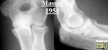 |
Mason Classification (1954)1. Undisplaced fracture (<2mm) 2. Marginal or rim fracture, displaced (original classification may be multifragmentary) 3. Multifragmentary fracture involving whole head |
Hotchkiss (1997), adaption of Mason
- 1.Undisplaced or minimally displaced (<2mm)
- 2. Displaced but amenable to internal fixation
- 3.Fracture that is not amenable to internal fixation
Treatment
Options
- 1. Symptomatic treatment and mobilisation (Aspirate and L/A most are thankful)
- 2. Open reduction and internal fixation
- 3. Radial head excision
- 4. Radial head excision and radial head prosthesis
When considering treatment consider extent of energy transfer. For displaced fractures type 2 and 3 aspirate and inject L/A. Assess ROM look for bony block and crepitus. Especially for type 3 fractures look closely clinically for evidence of injury to soft tissues. Notably:
- a. Interosseous ligament (membrane) in forearm ( forearm tenderness/ swelling)
- b. Wrist tenderness / DRUJ
- c. Medial collateral ligament of elbow (Risk instability if excise radial head)
- d. Look closely at coronoid (post dislocation of elbow with radial head injury)
Type 1
Symptomatic treatment and early movement. Some studies debating benefits of delaying mobilisation but mostly say mobilize as soon as possible.
Type 2
Aspirate and inject L/A
![]() (Click on image to see
how/ where to aspirate)
(Click on image to see
how/ where to aspirate)
- If no bony block and no crepitus, symptomatic treatment.
- If bony block or marked crepitus consider ORIF if fracture configuration suitable. ( Consider CT )
- If bony block / crepitus and not suitable for ORIF see type 3
Type 3
Treatment controversial and no ideal. Important to consider:
1. Functional demand
2. Extent of soft tissue injury and risk of instability. If excise head and evidence of interosseous ligament (membrane) damage, risk proximal migration of radius especially in high demand patient. If radial head fracture associated with post dislocation of elbow and or injury to medial collateral ligament risk instabilty if excise head without replacement.
Extremes 70 YO Type 3 with no forearm/ DRUJ pain best excise head early and mobilise. If 20 YO manual worker and evidence of significant soft tissue injury, work harder to preserve head or replace with prosthetic head.
Surgical approach to radial head
Classically the kocher interval is used, however several other approaches can be used.
See Approaches to elbow - Approaches
Direct links
References
Mason, M. L.. Some observations on fractures of the head of the radius with a review of one hundred cases. British J. Surg., 42: 123-132. 1954
Hotchkiss, R. N.. Displaced fractures of the radial head: internal fixation or excision?. J. Am. Acad. Orthop. Surgeons, 5: 1-10. 1997