Anterior approach to hip
For Incision and drainage - septic arthritis
Indication
Incision and drainage septic arthritis - paediatric
Classically the anterior approach to the hip is the Smith Peterson approach the full Smith Peterson approach begins on the iliac crest and extends down in the sartorius and tensor fascia interval. Similarly Salter described an approach to the paediatric hip for open reduction and innominate osteotomy.
These extensive approaches above are not required for drainage of a paediatric septic hip.
You still use the interval of Sartorius and Tensor fascia lata.
Anatomy
Lateral cutaneous nerve of thigh(
LCFN)
passes over sartorius about 2.5 cm distal to Anterior superior iliac spine
(adult). The
path of the LCFN is very variable see
LCFN.
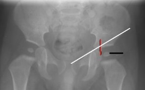
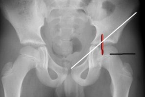
Mid inguinal point - This is NOT the
middle of the inguinal ligament but the mid point between the Anterior superior
iliac spine and pubic symphysis.
The mid inguinal point corresponds to the location of the common femoral artery (+- 1.5cm on either side in the adult Hunt et al).
The CFA overlies the femoral head. (Garrett)
Considerations
Classically the Smith Peterson approach is described as the anterior approach to the hip.
For incision and drainage of septic hip only a small portion/ window is required.
Positioning
Supine
Skin Incision
 |
Vertical or transverse (Suggest parallel to groin crease, pinch skin to see langer lines for more cosmetic scar )
Just below inguinal ligament overlying femoral head. (Use image intensifier if unsure to locate femoral head) You can often feel the interval between Sartorius and Tensor fascia lata. Place incision over the interval, essentially vertically down from ASIS.
Remember CVAN Canal Vein Artery Nerve Common femoral artery at mid inguinal point overlies head
|
 |
Superficial dissection
Develop interval between Sartorius and Tensor fascia muscle.
Mindful of lateral cutaneous nerve of thigh LCFN, identify the nerve and retract it medially with sartorius.
Deep to this is rectus femoris.
Mobilise rectus femoris medially exposing the hip capsule, beware of the ascending branch of lateral femoral circumflex artery which lies deep to rectus and sartorius and distal to hip joint.
Define a rectangle of capsule and excise a small window of capsule. (If unsure aspirate with a needle to confirm position, spend a little time defining capsule, makes it easier to excise window.)
Irrigate wound while moving hip around, place quill/ irrigation posteriorly through window, ensure you washout and suck out the posterior pocket of the hip capsule.
Consider suction drain into hip (remove 24 hours).
Closure
Muscles fall into place, simply close skin.
Post operatively
Obtain URGENT gram stain, ensure specimens plated out by microbiology immediately, don't leave till next day.
If true septic arthritis, consider abduction splinting to avoid hip subluxation.
Remove drain 24 hrs
Monitor clinical recovery, Temperature and inflammatory markers postop.
If definite septic arthritis consider placing double lumen PICC line for IV access and blood tests.
References
Garrett PD, Eckart RE, Bauch TD,
Thompson CM, Stajduhar KC; Fluoroscopic localization of the femoral head as a
landmark for common femoral artery cannulation. Catheter Cardiovasc Interv. 2005
Jun;65(2):205-7.
Hunt JA, Harris JP; Is the mid-inguinal point an accurate
landmark for the common femoral artery in vascular patients? Aust N Z J Surg.
1996 Jan;66(1):43-5.
Grothaus MC,
Holt M, Mekhail AO, Ebraheim NA, Yeasting RA; Lateral femoral cutaneous nerve:
an anatomic study. Clin Orthop Relat Res. 2005 Aug;(437):164-8.
Salter RB. Innominate osteotomy in the treament of congenital dislocation and subluxation of the hip. J Bone Joint Surg [Br] 1961;43-B:518–39 (PDF)
Personal observations