Postero lateral corner
Anatomy
 |
|
There is great variability in the anatomy and nomenclature of this region. Seebacher et al. divided the lateral part of the knee into three distinct layers.
- Lateral fascia, iliotibial tract, biceps tendon.
- Patellar retinaculum, patello-femoral ligament.
- Capsule, lateral collateral ligament (LCL), arcuate ligament, fabello-fibular ligament, popliteo-fibular ligament, tendon of popliteus.
The deep structures in detail:
The lateral collateral ligament
Arises from the lateral femoral condyle, in the mid-coronal plane, 2 cm above the joint line. It proceeds distally and posteriorly to the posterior aspect of the fibular head. The popliteus tendon courses beneath it. It is tubular in shape and is no more than 3 mm or 4 mm in diameter. The lateral ligament is superficial to and separate from the lateral capsule.The popliteus muscle and tendon
Arises from the postero-medial aspect of the shaft of the tibia above the soleal line. It has a number of distal attachments:
(i)The posterior horn of lateral meniscus via a muscular attachment. Its main insertion is to the lateral femoral condyle inferior to and passing underneath the lateral collateral ligament.
(ii)The posterior and middle segments of the lateral meniscus, via the inferior and superior popliteo-meniscal fascicles. These are the terminations of the popliteal tendon.
(iii)The apex of the fibula via the popliteo-fibular fascicle of the tendon.
Watanabe et al. found the popliteus to be present in all knees examined.
(iii). The popliteo-fibular ligament
This postage stamp sized structure is found deep to the lateral limb of the arcuate ligament. It arises from the posterior part of the fibula and inserts into the popliteus tendon proximal to its junction with the popliteus muscle belly. The popliteus tendon and popliteo-fibular ligament combined form an inverted Y-shaped musculo-tendinous structure with origins from the tibia and fibula with a common insertion on the lateral aspect of the femur. It is a major contributor to the strength of the posterolateral corner.
The arcuate ligament
This is a Y-shaped ligament which arises from the posterior part of the capsule around the distal surface of the femur and condenses down onto its insertion on the posterior aspect of the fibular head. In its course, it runs over the popliteus muscle, deep to the lateral inferior geniculate vessels. Variably present in 24% - 80% of knees.
The fabello-fibular ligament
The fabella is an inconsistent structure (sesamoid) present in the lateral head of gastrocnemius.The fabello-fibular ligament runs parallel to the lateral collateral ligament from the fabella to the fibula, inserting on the head of the fibula posterior to the insertion of the biceps tendon.
Biomechanics
Primary translations and rotations occur along the axis of the applied force or
moment, whereas coupled translations and rotations occur in directions different
to the applied force. For example, external tibial rotation that occurs in
response to a posterior force is called a coupled external rotation.
Selective sectioning of the ligaments revealed that:
If the lateral collateral or deep ligament complex (arcuate ligament, popliteus
tendon, fabello-fibular ligament and posterolateral part of the capsule) are cut
individually there is no change in posterior translation at all angles of knee
flexion. There is however an increase in varus translation, greater with LCL
section than the deep ligament complex.
If the LCL and deep ligament complex are cut together, there is a slight
increase in posterior translation at all angles of flexion and an increase in
the coupled external rotation with posterior force at all angles of knee
flexion. Also, there is an increase in varus rotation in response to a varus
force; this is maximal at 30° of knee flexion and is significantly greater than
if the LCL alone is cut.
If the posterior cruciate ligament (PCL) alone is cut there is increased
posterior translation at all angles of flexion, increasing as the knee is flexed
from 0° to 90°. Also, there is a cessation of coupled external rotation with
posterior force.
If the PCL, LCL and deep ligament complex are all cut then there is a much
greater posterior translation at all angles of flexion, a greater varus rotation
in response to varus force, maximal at 60° of knee flexion and an increase in
primary external rotation.
In essence, section of posterolateral structures results in increased primary
posterior translation, primary varus rotation, primary external rotation and
coupled external rotation.
La Prade et al. have measured the forces acting upon anterior and posterior
cruciate ligament grafts following section of posterolateral structures. In both
grafts, forces increased with varus loading and even more with coupled varus and
external rotation.
Mechanism of injury to the posterolateral corner
Most likely combined hyperextension and varus force. eg. posteromedially angled blow to an extended leg.
Other mechanisms include coupled hyperextension and external
rotation of the tibia, tibial external rotation force and a heavy varus force.
The posterolateral structures are also frequently damaged in severe knee
injuries such as complete dislocation.
Posterolateral corner injuries in isolation do exist but in most instances the
posterolateral structures are disrupted in combination with the PCL (most
commonly), anterior cruciate ligament (ACL) or both.
Diagnosis
History
Acute or chronic
Acutely patients present with swelling, posterolateral knee pain and
occasionally numbness and weakness of the foot due to peroneal nerve
symptoms. As the acute swelling improves, the patient may notice
instability of the extended knee, which may abnormally hyperextend as weight is
put through it with walking.
With more chronic injuries, the presentation may be with lateral or medial joint
line pain, persistent peroneal symptoms or instability. Again, this instability
usually causes hyperextension problems, frequently noticed when ascending or
descending stairs. The instability may also be noticed on external rotatory
movements which cause posterior subluxation of the lateral tibial plateau. This
movement was termed posterolateral rotatory instability by Hughston et al.
Instability may evolve a while after the injury and may be confounded by
concomitant injuries to the cruciate ligaments. It is often very difficult to
distinguish isolated cruciate injuries from those complicated by posterolateral
corner injuries.
Examination
Several tests and examination points have been described:
Gait
Acutely - Antalgic gait
In more chronic cases during the stance phase of gait patients may have a varus thrust or hyperextension varus thrust of the injured knee. It has been postulated that this is due to external rotation of the tibia in full extension causing an apparent tibia vara. Some patients may walk with a slightly flexed knee, this may be to avoid the pain and instability they experience in hyperextension or to avoid stresses on the joint and capsule which are greater in hyperextension. When standing patients may be noticed to have a varus alignment of the knee.
Look
Look for swelling, bruising
Feel
Palpate the posterolateral joint line looking for tenderness.
Anterior/posterior translation
In all tests of ligament function comparison is made with the uninjured knee.
-
Anterior posterior translation tested at 30° and 90° of knee flexion.
-
Increased posterior translation at 30° only indicates posterolateral injury, whereas at both 30° and 90° it indicates injury of the PCL.
-
ACL integrity should be assessed with the Lachman test; there may however be a slight anterior translation if there is an isolated posterolateral injury (this though will have a firm end-point if the ACL is intact).
Dial test (tibial external rotation test)
-
Patient prone or supine
-
Knee flexed to 30° and to 90°
-
Grasp the soles of the feet and apply an external rotation force
-
The medial border of the foot is used as the reference point against the axis of the femur
-
Look for a difference in external rotation between each leg revealed as an increase in thigh foot angle.
-
It is important that the measurement is a comparative one as there is a wide variation in the amount of rotation in normal knees.
-
> 10° increase in external rotation compared to contra lateral side is pathological.
Posterolateral drawer test
As described by Hughston and Norwood.
-
Patient supine
-
Hip flexed to 45°, the knee flexed to 80° and the tibia in 15° of external rotation.
-
The foot is fixed and a posterior force applied to the tibia.
-
Positive for injuries to the posterolateral corner when the lateral tibial condyle rotates relative to the lateral femoral condyle. Again compare with contralateral side.
-
If the posterolateral drawer is grossly positive then injury to the PCL and posterolateral corner should be suspected.
External rotation recuvartum test
As described by Hughston and Norwood demonstrates posterolateral instability in the extended knee.
-
Patient supine
-
Lift both legs by grasping the big toes
-
Compare for differences in: lateral hyperextension, varus and tibial external rotation.
Imaging
Radiographs
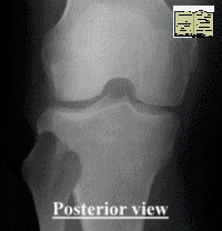
Features on X-ray suggestive of posterolateral injury include:
-
segond fracture (bone avulsion of capsule from mid-third lateral tibial plateau)
-
abnormal widening of lateral joint space
- avulsion fracture of fibula head
- avulsion fracture of Gerdy's tubercle.
These are however all non-specific findings and may be associated with isolated ACL or PCL injury.
MRI
Preferred imaging modality.
Examination under anaesthesia and arthroscopy
EUA above tests/ signs
Arthroscopy allows
visualisation and confirmation of associated cruciate injuries and allows
diagnosis of meniscal and chondral pathology which may have been occult on MRI.
Arthroscopy can also
be used to visualise the popliteal hiatus and assess damage to the popliteus
complex.
Treatment
Treatment slightly controversial as a whole acute repair is better than delayed reconstruction.
Isolated low-grade injuries to the posterolateral corner may do well with conservative treatment.
In small studies, non-operative treatment of what were deemed mild to moderate
injuries to the posterolateral corner have had a good outcome but in more severe
injuries, the outcome is poor.
It is however very rare to get an isolated injury to the posterolateral corner,
as such injuries are nearly always associated with injury to the ACL, PCL or
both. It is now advised that in such multiple injuries that all elements of the
instability should be surgically addressed for a good outcome to be attained.
O'Brien et al. showed that failure of ACL reconstruction can often be due to
unrecognised and hence untreated posterolateral corner deficiency.
The aim of surgery is to correct pathologic instability by correcting the
abnormal external tibial rotation and varus instability caused by the
posterolateral insufficiency, whilst the antero-posterior instability is
addressed by ACL or PCL reconstruction.
Several reconstruction options exist, below is one.
Principles of repair
With an acute injury individual
structures can be directly repaired. This requires early exploration (within 72
h) and a very detailed knowledge of the anatomy of the region.
With chronic injuries it is impossible to repair individual structures, and thus
the goal is therefore to fashion a reconstruction that mimics the function of
damaged structures.
Restore bony alignment. A valgus high tibial osteotomy may be needed in addition
to reconstruction in cases of severe varus deformity to prevent excessive force
being placed on the lateral capsular structures being reconstructed.
A strong soft tissue post of either allograft or autograft is needed to support
the posterolateral corner, either as the reconstruction or to support any
primary repair of tissues.
Any reconstructed post should be isometric through all angles of knee flexion
and extension.
A technique of repair
Larson technique using autogenous semitendinosus free graft to reconstruct the
LCL and popliteo-fibular ligament. This is most frequently performed in
combination with an ACL or PCL reconstruction.
1. Semitendinosus graft is harvested from the ipsilateral limb (or the
contralateral limb if the ipsilateral tissues are used for concomitant ACL/PCL
reconstruction).
2. The posterolateral aspect of the leg is exposed via a curvilinear incision,
exposing the fibular head and lateral femoral condyle.
3. A tunnel is created in the fibular head in a lateral direction, initially
with a guide wire, then widened with a drill. The tendon graft is passed through
the fibular head drill hole.
4. The free ends of the graft are passed beneath the iliotibial band and biceps
tendon.
5. A guide pin is used on the femoral condyle to check for isometry by looping
the ends of the graft around it. Once a satisfactory position has been found,
the guide wire is passed through the femur to penetrate the medial aspect of the
knee. A blind-ending 30 mm tunnel is drilled over the guide pin on the lateral
side.
6. The ends of the graft are passed through the tunnel via a passing suture and
tensioned by pulling on the passing suture. The graft is fixed via a
bioabsorbable interference screw 1–3 mm bigger than the diameter of the tunnel
Summary
Injuries to the posterolateral
structures of the knee are common accompaniments to ACL and PCL rupture;
isolated posterolateral injury is rare. It is important to recognise and treat
posterolateral injuries as failure to do so when reconstructing cruciate
ligaments can result in failure.
Posterolateral injury usually manifests as pathologic external tibial rotation
and varus instability. It is best detected with the dial and posterolateral
drawer tests on examination. Diagnosis can be aided with MRI and arthroscopy.
Surgery should aim to address all elements of the instability and should allow
the patient to return to a near normal level of function although combined
injuries are often a major insult to the knee and may never fully recover
despite appropriate and timely surgical reconstruction.
References
The posterolateral corner of the knee: Anatomy, biomechanics and management of injuries, Injury, Volume 35, Issue 1, January 2004, Pages 68-75; Hywel Davies, Andrew Unwin and Paul Aichroth