Ankle Overview
The secret to assessment of ankle injuries/ radiographs is "The mortise".
The Talus is supported in the mortise by medial and lateral columns.
These columns consist of two elements -
- Bone ( medial and lateral malleoli)
- Ligaments. (Medial and Lateral colateral ligaments)
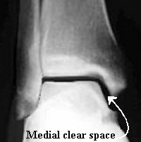
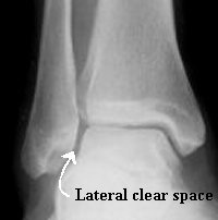
It is important that the Talus is congruent in the mortise. The medial clear space should be within 1-2 mm of the superior clear space. The Lateral clear space changes with rotation of the leg as the fibula sits in a groove postero-lateral to the tibia. If you suspect that that the Talus is not congruent in the mortise ask for a mortise view. (see below) This is taken with the foot/ ankle in 15 - 20° of internal rotation. This brings the Fibula around out to the side of the Tibia, allowing you to asses the lateral clear space.
If you see the Talus has shifted and is not congruent in the mortise, work out what has broken.
For the talus to shift you have to injure something on both sides of the mortise (bony or ligamentous).
Radiographs
 |
 |
 |
AP viewFibula sits slightly posterior to Tibia therefore on Ap view no lateral clear space visible. Medial clear space within 1-2mm of superior clear space. |
Mortise ViewTaken with foot/ankle 15 Degrees of internal rotation, now see lateral clear space. Allows you to assess congruence of mortise and look for evidence of Talar shift |
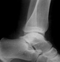
Lateral ViewLook at posterior malleolus |