Ankle Fractures
Anatomy and Biomechanics
The articulation between Tibia and dome of the Talus is a highly congruent saddle-shaped weight-bearing surface.
The medial talar facet articulates with the medial malleolus and the lateral facet articulates with the lateral malleolus.
During weight-bearing, 80 to 90% of the load is transmitted through the tibial plafond to the dome of the talus. With varus and valgus stress, as much as 22% of the load can be transmitted through the medial facet and as much as 10% through the lateral facet.
Under normal circumstances, 17% of the total load is transmitted proximally through the fibula, although the mechanism by which the load is transmitted to the fibula through the syndesmosis is not fully understood.
The fibula is bound to the tibia by the anterior and posterior tibiofibular ligaments and the syndesmotic ligament, which begins between the tibiofibular ligaments and extends proximally between the tibia and the fibula.
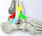
The lateral collateral ligament
The lateral collateral ligament is made up of three separate ligaments.
|
|
|
Medial collateral ligament (Deltoid ligament)
Two layers
-
Superficial - origin anterior and inferior aspect of the medial malleolus, insertion talar neck and calcaneus
-
Deep - origin inferior and posterior aspect of the medial malleolus, insertion medial and posteromedial aspects of the talus
Although the ankle joint has been thought of as a hinged articulation in the past, it has been demonstrated that the normal motion of plantar flexion and dorsiflexion is a combination of sliding and rolling.
The normal range of motion of the ankle ranges from 12º of dorsiflexion to 56º of plantar flexion in the unloaded state.
Plantar flexion causes between 4 and 8º of internal rotation of the talus; this rotation is thought to be a consequence of tethering of the medial aspect of the talus by the deltoid ligament.
The motion of the talus in plantar flexion and dorsiflexion causes motion of the fibula. As the ankle dorsiflexes, the fibula moves in a posterior-to-lateral direction and externally rotates as much as 2º. There does not appear to be any major vertical component to this motion.
Although plain radiographs have suggested that when there is instability of the ankle joint the primary alteration is caused by a lateral shift of the talus, this is a visual illusion created by a two-dimensional representation of a three-dimensional situation. Several investigators have demonstrated that the primary pattern of instability is external rotation of the talus. This finding is consistent with the observation that sectioning of the anterior tibiofibular ligament does not by itself cause talar instability, while sectioning of the deep deltoid ligament leads to talar instability. Clinically, it has been observed that the absence of a medial malleolus or a displaced pseudarthrosis of the medial malleolus can lead to osteoarthrosis, although frank instability is not usually seen. These data suggest that the primary stabilizing structure of the ankle is on the medial rather than on the lateral side, in contrast with the conclusions of earlier reports.
Under normal physiological conditions, the ankle joint sustains peak loads of almost four times body weight. This loading actually serves as a stabilizing influence on the joint because it causes the talus to seek an anatomically reduced position underneath the tibial plafond (by means of an associated two-millimeter lateral talar shift). This acts to reduce motion of the injured ankle. The importance of the axial load in experimental testing is apparent from the observation that loading increases the joint-contact area by as much as 100% resulting in increased stiffness and altered kinematics. Consequently, conclusions regarding the stability of the joint that have been based on experiments performed on unloaded specimens of the ankle cannot be extrapolated to normal conditions.
Radiographs
The standard radiographs to asses an ankle are a Mortise and lateral view.
Different values have been suggested for the significance of the medial clear space.
Some have suggested > 1mm, compared to the superior clear space. Some have suggested > 4 or 4.5 mm.
Saldua et al showed that plantar flexion of the foot does effect the width of the medial clear space but only by around 0.38mm. It does affect it enough though to lead to some false positives for deltoid ligament injury.
If unsure about medial (deltoid ligament injury) consider gravity external rotation stress views. Gravity external rotation stress views are as good as manual stress radiographs. Gill et al
Aims of Treatment
The aim of treatment
is to restore biomechanical stability to the ankle. Clearly, an isolated fracture of the lateral malleolus is
biomechanically distinct from a bimalleolar fracture.
There are two indications for the operative treatment of a fracture about the
ankle.
-
Static incongruity - a step-off in a weight-bearing portion of the articular surface. This rarely occurs in fractures about the ankle, except in pilon fractures, so it will not be discussed further.
-
Dynamic incongruity - instability, abnormal tracking of the talus within the ankle.
The main risk of operative intervention is infection 1- 4%.
Classification
Descriptive
- Medial Malleolus
- Lateral Malleolus
- Bimalleolar - Both medial and lateral Malleolus
- Tri malleolar - Medial, Lateral, and Posterior malleolus
Lauge Hansen (1950)
The rationale for the development of the Lauge-Hansen classification was to guide the closed treatment of fractures about the ankle. Don't learn by heart, try understand principles and work out how it happens.
Looks at two things:
-
Position of foot - Supination or Pronation
-
Direction of deforming force - External rotation, Abduction, Adduction
The position of the foot at the time of injury determines which structures are taught or under tension.
The injury begins on the side where the structures are already under tension, then moves around the ankle with further structures failing sequentially.
-
Supination, External rotation (SER)
With the foot supinated the deltoid (medial) ligament is relaxed therefore the injury begins laterally. With external rotation the anterior Tibiofibular ligament fails first, if the force continues the fibula fails in a short oblique/ spiral fracture, if it continues further the posterior tibiofibular ligament fails and finally if the force continues the medial malleolus fails.
-
Supination, Adduction injury (SA)
Again the Deltoid ligamentt is relaxed, so the injury begins laterally. With Adduction and no rotational force the Lateral colateral ligament (ATFL, CFL and PTFL) fails or pulls off the tip of the lateral malleolus (transverse fracture), if the force continues it pushes of the medial malleolous.
-
Pronation, External rotation (PER)
In pronation the Deltoid ligament is taught. Therefore the injury begins medially, rupturing the deltoid ligament or pulling of the medial malleolus. If the force continues the rotational force then rotates the fibula away from the tibia, rupturing the anterior Tibio fibula ligaments, if the force continues the fibula fails (high) and then the posterior tibiofibular ligaments.
-
Pronation, Abduction (PA)
In pronation the Deltoid is taught. Therefore the injury begins medially, rupturing the deltoid or pulling off the medial malleolus. If the force continues laterally (pushing off) breaking the lateral malleolus.
Weber
Looks at the level of the fracture in the lateral malleolus, medial injuries are not considered.
-
Type A - Distal to the joint line
-
Type B - Begins at the joint line and follows a spiral, oblique, upward direction
-
Type C - Proximal to the level of the syndesmosis, ie injury involves the syndesmosis.
The problem with the Weber system is, a type-B fracture of the lateral malleolus without an associated medial injury behaves differently, from a biomechanical perspective, than a similar fracture that is accompanied by a fracture of the medial malleolus.
However, both types of fracture are classified identically according to the Weber system.
The inter-observer reliability for both systems is poor to fair, with less than 60% agreement among viewers regardless of their level of experience.
Isolated Fractures of the Lateral Malleolus
Up to 85% of lateral malleolar fractures occur without substantial injury to the medial side.
Lateral displacement of the talus decreases the tibiotalar surface contact area by 42% remember its not a flat surface but sadle shaped. This is considered to be evidence of the importance of the lateral malleolus in the maintenance of stability of the ankle. However, the model for this investigation was non-physiological since it involved the placement of spacers between the talus and the medial malleolus in order to force the talus laterally. Yablon et al demonstrated, experimentally and clinically, that the position of the talus in an ankle that has a bimalleolar fracture is not acceptable until the lateral malleolus has been anatomically reduced. This led to the concept that the talus faithfully followed the lateral malleolus.
This conclusion is applicable to bimalleolar fractures of the ankle but not to isolated fractures of the lateral malleolus. As important as these investigations were to the development of an understanding of fractures about the ankle, they were both limited by the biomechanical testing apparatus available at the time that the studies were performed. Since the ankle joint is not a hinge but a complex joint with coupled motions, the testing of ankles with use of highly constrained systems that do not provide for this motion will not yield physiologically relevant data. More recent studies that have provided for unconstrained motion of the ankle during testing have uniformly demonstrated that an isolated lateral injury about the ankle does not lead to abnormal mechanics or kinematics of the joint.
It has also been shown that the amount of displacement of the fibula
does not determine talar displacement when the ankle is axially loaded.
Clinical studies limited to isolated fractures of the lateral
malleolus have uniformly shown no advantage for operative treatment compared
with closed treatment, even when there was as much as
3mm of fibular displacement.
The concept that closed, non-operative treatment of isolated fractures of the
lateral malleolus can yield satisfactory results is based on a biomechanical
understanding of these fractures. In the absence of a medial injury,
dynamic incongruity cannot be demonstrated experimentally, implying that the
apparent displacement of the lateral malleolus is not clinically important.
This concept was supported by a computed tomographic investigation in which the apparent external-rotation deformity of the lateral malleolus was actually found to represent internal rotation of the proximal aspect of the fibular shaft that had occurred after the fracture.
Talocrural angle, indirectly measures proximal shortening of the fibula. This is the angle created by the intersection of a line drawn through the distal parts of the malleoli and a line bisecting the tibial shaft. Clinically important shortening is ascertained by a difference of between 2 and 5º in the talocrural angle compared with that in the contralateral, normal ankle.
In a recent study, this angle was measured on plain radiographs and with the use of three-dimensional computed tomography. It was found that the angle could not be used to distinguish between fractures that necessitated operative treatment because of the position of the fibula and those that did not need such treatment.
Combined Medial and Lateral Fractures
A bimalleolar fracture about the ankle involves a lateral malleolar fracture and either a medial malleolar fracture or a rupture of the deltoid ligament.
Experimentally, either type of medial injury has resulted in an equivalent change in the tibiotalar contact area or in an alteration in the kinematic behavior of the joint.
Clinical studies have shown improved outcome following operative intervention for bi malleolar fractures. An anatomical reduction was the primary determinant of a successful outcome.
One of the most controversial questions with respect to bimalleolar fractures involves the criteria for the diagnosis of a clinically and biomechanically important injury of the deltoid ligament in the absence of a medial malleolar fracture. A disruption of the deltoid ligament can be diagnosed with relative confidence when the medial clear space between the talus and the medial malleolus is increased.
If, in the presence of medial tenderness, more than 5mm of space is seen either initially or on a stress radiograph, a presumptive diagnosis of a substantial injury of the deltoid ligament can be made.
Such injuries should be treated as bimalleolar fractures, with open reduction and internal fixation of the lateral malleolus.
Routine exploration of the medial side of the ankle is not necessary unless there is evidence that a portion of the deltoid ligament has entered the joint and is blocking reduction of the talus.
Open Injuries
Gustilo and Anderson grade-I, II, or IIIA open fractures about the ankle should be treated with immediate internal fixation and delayed closure.
The more severe (grade-IIIB and IIIC) fractures should be treated with debridement and external fixation.
Injuries of the Syndesmosis
Fibular fractures that begin proximal to the tibial plafond are assumed to include an injury of the syndesmosis. Until recently, this injury was thought to be an absolute indication for fixation of the syndesmosis.
However, Boden et al showed, in a cadaver model, that, in the absence of a medial injury, no amount of disruption of the syndesmosis altered the loading characteristics of the ankle. In the presence of a medial injury, disruption of the syndesmosis that extended more than 4.5 cm proximal to the joint line did affect the loading characteristics of the ankle; with less than 3 cm of disruption, no alteration was seen. At between 3 and 4.5 cm of disruption, inconsistent results were reported.
Measurements of intra-articular pressures in the ankle have also shown that complete disruption of the syndesmosis does not affect pressures until the deltoid ligament has been transected. At this point, the tibiotalar contact area has decreased by 40% and the contact pressures have increased by 36%. The syndesmosis has not appeared to widen until the medial side has been disrupted, again indicating the importance of the medial structures in the maintenance of stability of the ankle.
The diagnosis of widening of
the syndesmosis is made when there is a space of more than 5 mm
between the distal aspects of the tibia and the fibula, as seen on the mortise
radiograph. Allowing a persistent displacement of the fibula would lead to a
poor clinical outcome.
The results of operative treatment of syndesmotic injuries can be heavily
influenced by the specific operative techniques that are used. A screw that is
placed from the fibula medially into the tibia should engage 3 or 4
cortices and should be 4.5 mm in diameter.
Fixation with a lag screw should not be performed since this could result in over tightening of the screw and narrowing of the syndesmosis. If this happens, dorsiflexion of the ankle will be diminished.
In addition, the screw should be parallel to the joint to avoid displacement of the fibula in an inferior or superior direction. Although there has been some interest in the use of intramedullary rods for the fixation of fibular fractures, it should be noted that, in the context of a syndesmotic injury, this method will make reduction and stabilization of the syndesmosis very difficult.
Controversy persists regarding removal of the syndesmotic screw before allowing full weight-bearing postoperatively.
Although several authors have advocated removal of the syndesmotic screw before weight-bearing, there have been few reports of breakage of screws that have been left in place.
The screw does not alter the range of dorsiflexion or of plantar flexion of the ankle and, if the screw engages only three cortices, the normal external rotation of the fibula during dorsiflexion of the ankle will not be affected. Radiolucency has been noted around such screws after patients have started to bear weight, suggesting that loosening of the screw permits normal motion of the fibula.
Removal of the screw involves some risk since this procedure, if done 6 weeks postoperatively, can be accompanied by redisplacement of the fibula.
Posterior Malleolar Fractures
The fracture fragment represents an avulsion by the posterior tibiofibular ligament at its site of attachment to the tibia.
The clinical results associated with these fractures are worse than those associated with bimalleolar fractures, particularly when the injury involves large fragments of the tibial plafond.
Experimentally, fractures of more than 30% of the posterior aspect of the tibia have led to instability of the ankle when tested in a posterior direction. Reduction and fixation of the fibular fracture frequently reduces the fracture of the posterior malleolus. This decreases the tibiotalar instability, even in the absence of direct fixation of the posterior malleolar fragment.
McDaniel and Wilson showed that closed reduction of fractures involving less than 25% of the posterior tibial surface led to a good or excellent result in 18 of 28 patients, even in the presence of residual displacement of more than 2 mm. If the fragment comprises more than 25% of the surface, according to estimates of the fracture size made from plain radiographs, a good or excellent result can be expected in only 1 of 4 patients.
Open reduction and fixation of the posterior malleolus can be accomplished with use of a screw, directed either anteriorly or posteriorly. A stab incision can be made over the tibia from the anterior direction, and a lag screw can be placed to engage the posterior fragment. This fragment will have been reduced digitally and held in place with use of a lateral incision. Alternatively, the posterior fragment can be approached directly through a posterolateral incision. This permits reduction of the fragment under direct visualization and allows placement of a lag screw from a posterior-to-anterior direction. Although the latter approach involves more dissection, it can result in a more reliable reduction and stabilization of the posterior fragment.
Postoperative Management
Several authors have advocated early motion after operative treatment of fractures about the ankle. This recommendation is based on the experimental observation that continuous passive motion in rabbits decreased the stiffness of the joint. Although one study indicated that early motion with weight-bearing yielded better motion of the ankle after 3 months, most investigators have been unable to demonstrate any substantial difference in the range of motion or the level of activity of the patient between ankles that had been treated with early motion and those that had been immobilized continuously for 3 to 6 weeks.
Non-operative treatment may be followed by a slightly faster recovery of the range of motion, but no lasting advantage has been shown.
Similarly, it has been demonstrated that weight-bearing affects neither the time to recuperation nor the clinical result in either operatively or non-operatively treated fractures of the ankle.
Moreover, there is no evidence that any specifically designed rehabilitation program for the treatment of healed fractures after immobilization has any effect on the clinical result.
In the absence of subsequent trauma, virtually all fractures about the ankle will have osseous union.
A good or excellent result can be expected after treatment of 85 to 90% of displaced bimalleolar fractures if an anatomical reduction has been accomplished operatively.
If a
non-anatomical reduction has been obtained, the results tend to be poorer; only
60 to 80% good and excellent results have been reported. In one report, it was suggested that the clinical
result is not only a function of an anatomical reduction; it also depends on the
presence of a concomitant talar injury.
Patients can expect a gradual improvement in the function of the ankle as late
as 9 years postoperatively. Osteoarthrosis, when it occurs, usually does so
within the first 2 years after the injury. Radiographic evidence of
degeneration of the articular cartilage is seen in association with less than 5% of unimalleolar fractures and with as many as 20% of bimalleolar
fractures. The degeneration, however, does not have a strong association with
pain in the ankle that limits function.
Swelling is a universal finding after a fracture of the ankle, but it is more severe and more persistent after operative treatment.
The size of the ankle tends to return to normal 3 months after closed treatment and 9 months after operative treatment .
However, in one series, 24 (49%) of 49 patients reported intermittent swelling of the ankle as late as 18 months postoperatively.
A malunion of an ankle fracture sometimes presents with persistent swelling more than 2 years after the injury.
A malunion of the fibula may be fairly subtle, and computed tomography may be needed to delineate the aberrant anatomy.
A fibular malunion decreases tibiotalar contact by approximately 30%, which would be expected to increase contact pressures.
Metalwork removal
Metal work removal is not associated improved results or a reduction in long-term complications.
In one study, 59 (89%) of 66 patients who had hardware removed did so because of discomfort.
Although relief of local hardware-related discomfort can be expected in almost all such patients, removal of the hardware necessitates a second operative procedure with its attendant risks and costs.
Biodegradable fracture-fixation implants theoretically reduce the need for this second operation. Polyglycolide (Dexon) has been used widely as a suture, most of the polymer that is in orthopaedic use has been associated with the formation of sterile abscesses (in as many as 11% of patients), with as many as 5% of such abscesses necessitating a second operation.
Furthermore, this polymer may not possess adequate strength, as indicated by a rate of mechanical failure of as high as 5% (2 of 41 patients). In a recent prospective, randomized study, more encouraging results were reported with use of 4.0-millimeter polylactide screws for fixation of the medial malleolus.
At the 3-year follow-up evaluation, the radiographic and functional results were equivalent to those obtained after use of stainless-steel screws, and no sterile abscesses formed.
There is continuing research in many centres around the world for further development of orthopaedically useful biodegradable implants.
References
Current Concepts Review. Fractures about the Ankle; JBJS A 77 January 1995 pg142-152; Michelson, James D.
Plantar flexion influences radiographic measurements of the ankle mortise. J Bone Joint Surg Am. 2010 Apr;92(4):911-5. Saldua NS, Harris JF, LeClere LE, Girard PJ, Carney JR.
Comparison of manual and gravity stress radiographs for the evaluation of supination-external rotation fibular fractures J Bone Joint Surg Am. 2007 May;89(5):994-9. . Gill JB, Risko T, Raducan V, Grimes JS, Schutt RC Jr.