© Cambridge Orthopaedics - Cambridge; United Kingdom


Elbow Anatomy
To understand the anatomy of the elbow it is important to understand a few medical terms
•
Flexion - Bending your elbow, as in bringing your hand to your mouth
•
Extension - Straightening your elbow
•
Supination - Twisting your forearm and hand so your palm faces the ceiling (asking for change)
•
Pronation - Twisting your forearm and hand so the back of your hand faces the ceiling
•
Lateral - Outside of your arm/ elbow (furthest from the midline of the body)
•
Medial - Inside of your arm/ elbow (nearest the midline of the body)
The elbow is more than a hinge joint, allowing for bending the arm/ elbow, flexion and extension. It also allows for rotation of the forearm wrist and
hand, supination and pronation. It is made up of:
•
Muscles and tendons
•
Ligaments
•
Bones
•
Nerves
•
Blood vessels
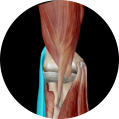
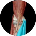
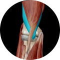
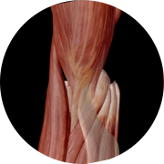
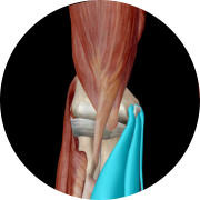
Muscles and tendons
Tendons connect muscles to bone and transfer all the force generated by the muscles. All the muscles that extend your
wrist and fingers attach to a small bony area on the outer side (lateral side) of your elbow, otherwise called the
common extensor origin. It is pain here that is called tennis elbow (lateral epicondylitis).
All the muscles that flex your wrist and fingers attach to a small area of bone the medial epicondyle on the inner side
of your elbow. It is pain here that suggests golfers elbow (medial epicondylitis).
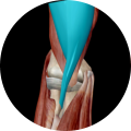
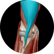
Several muscles work to flex the elbow, most people think the biceps muscle is the main flexor of the elbow, in fact
Brachialis a muscle beneath biceps is the main elbow flexor. Biceps is very important for supination (asking for change).
Triceps is the main muscle straightening the elbow (extension).
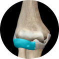
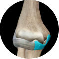
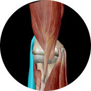
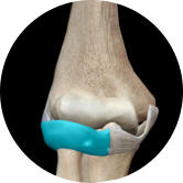
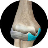
Ligaments
Ligaments connect bones to bones, they help stabilise the elbow, stopping it from dislocating. There are essentially three ligament complexes around
the elbow.
•
Medial collateral ligaments
•
Lateral collateral ligaments
•
Annular ligament
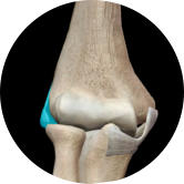
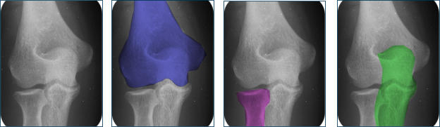
Bones
The elbow joint is made up 3 bones:
•
Humerus
•
Radius (Radial head)
•
Ulna (Olecranon)
The bone making up your upper arm is the
humerus, the humerus connects to the forearm bones at the elbow joint. The forearm contains two bones, the radius and the ulna.
The radius runs from the outer side of your elbow down the thumb side of your forearm. The ulna is on the inner side.
The elbow joint has essentially two joints within it - one a hinge joint that allows bending and straightening of the elbow, the other allows for rotation
or twisting. It allows the forearm to twist (pronate and supinate) so you can show the back of your hand and twist your forearm to ask for change.
The place where the ulna joins the humerus is called the ulna humeral joint and this is essentially the hinge joint that allow for bending and
straightening of the arm.
The radius ends in the radial head at the elbow, it joins onto the humerus at the elbow. The small part of the joint on the humerus side where the
radius attaches to it is called the capitellum and as such this half of the elbow joint is called the radiocapitellar joint. The radiocapitellar joint allows
for rotation of the forearm, wrist and hand (pronation and supination).
In the elbow joint the ends of the bones are covered in articular cartilage, this is a very smooth slick material that allows the joint surfaces to slide
over each other.
Subchondral bone is a layer of hard bone that lies below the articular cartilage and supports the joint and anchors the cartilage onto the bone.
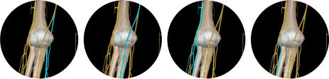
Nerves
There are three main nerves that cross the elbow and several small nerves that cross the elbow providing sensation to the skin on the forearm.
The three main nerves are:
•
Ulna nerve
•
Median nerve
•
Radial nerve (the PIN, posterior interosseous nerve is a branch)
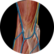
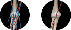
Blood vessels
The main vessel above the elbow is the brachial artery just below the elbow it divides into two the ulnar and the radial artery.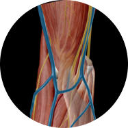


© Cambridge Orthopaedics - Cambridge; United Kingdom
© Cambridge Elbow - Cambridge; United Kingdom
Elbow Anatomy
To understand the anatomy of the elbow it is important to understand a
few medical terms
•
Flexion - Bending your elbow, as in bringing your hand to your
mouth
•
Extension - Straightening your elbow
•
Supination - Twisting your forearm and hand so your palm faces the
ceiling (asking for change)
•
Pronation - Twisting your forearm and hand so the back of your
hand faces the ceiling
•
Lateral - Outside of your arm/ elbow (furthest from the midline of
the body)
•
Medial - Inside of your arm/ elbow (nearest the midline of the body)
The elbow is more than a hinge joint, allowing for bending the arm/ elbow,
flexion and extension. It also allows for rotation of the forearm wrist and
hand, supination and pronation. It is made up of:
•
Muscles and tendons
•
Ligaments
•
Bones
•
Nerves
•
Blood vessels
Muscles and tendons
Tendons connect muscles to bone and transfer all the
force generated by the muscles. All the muscles that
extend your wrist and fingers attach to a small bony
area on the outer side (lateral side) of your elbow,
otherwise called the common extensor origin. It is pain
here that is called tennis elbow (lateral epicondylitis).
All the muscles that flex your wrist and fingers attach to
a small area of bone the medial epicondyle on the inner
side of your elbow. It is pain here that suggests golfers
elbow (medial epicondylitis).
Several muscles work to flex the elbow, most people think
the biceps muscle is the main flexor of the elbow, in fact
Brachialis a muscle beneath biceps is the main elbow
flexor. Biceps is very important for supination (asking
for change).
Triceps is the main muscle straightening the elbow
(extension).
Ligaments
Ligaments connect bones to bones, they help stabilise the
elbow, stopping it from dislocating. There are essentially three ligament
complexes around the elbow.
•
Medial collateral ligaments
•
Lateral collateral ligaments
•
Annular ligament
Bones
The elbow joint is made up 3 bones:
•
Humerus
•
Radius (Radial head)
•
Ulna (Olecranon)
The bone making up your upper arm is the humerus,
the humerus connects to the forearm bones at the
elbow joint. The forearm contains two bones, the
radius and the ulna.
The radius runs from the outer side of your elbow
down the thumb side of your forearm. The ulna is on
the inner side.
The elbow joint has essentially two joints within it -
one a hinge joint that allows bending and
straightening of the elbow, the other allows for
rotation or twisting. It allows the forearm to twist
(pronate and supinate) so you can show the back of
your hand and twist your forearm to ask for change.
The place where the ulna joins the humerus is called
the ulna humeral joint and this is essentially the
hinge joint that allow for bending and straightening
of the arm.
The radius ends in the radial head at the elbow, it
joins onto the humerus at the elbow. The small part
of the joint on the humerus side where the radius
attaches to it is called the capitellum and as such this
half of the elbow joint is called the radiocapitellar
joint. The radiocapitellar joint allows for rotation of
the forearm, wrist and hand (pronation and
supination).
In the elbow joint the ends of the bones are covered
in articular cartilage, this is a very smooth slick
material that allows the joint surfaces to slide over
each other.
Subchondral bone is a layer of hard bone that lies below the articular
cartilage and supports the joint and anchors the cartilage onto the bone.
Nerves
There are three main nerves that cross the elbow and several small nerves
that cross the elbow providing sensation to the skin on the forearm.
The three main nerves are:
•
Ulna nerve
•
Median nerve
•
Radial nerve (the PIN, posterior interosseous nerve is a branch)
Blood vessels
The main vessel above the elbow is the brachial artery just below the elbow it divides into two the ulnar and the radial artery.