Sciatic nerve block
Sciatic nerve:
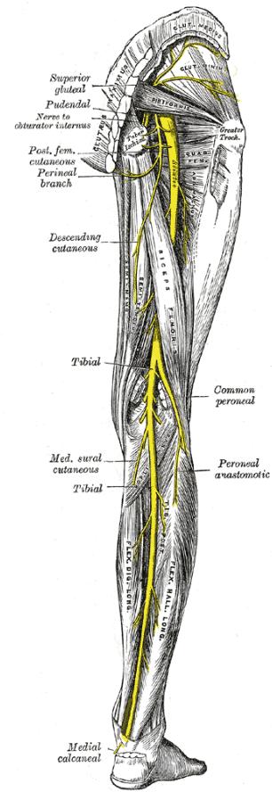
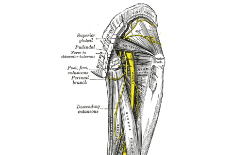
It originates from ventral rami of L4-S3.It exits the pelvis through greater sciatic foramen underneath
the piriformis muscle.
It then passes between gluteus maximus and quadratus femoris muscle lying
in between the greater trochanter and ischial tuberosity.
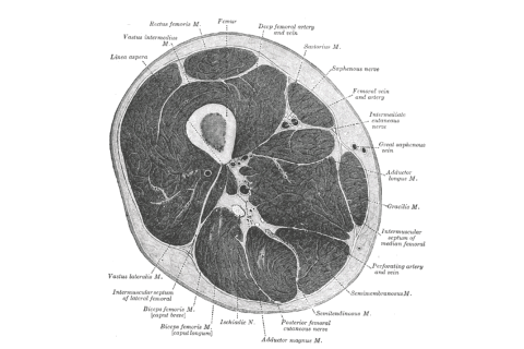
In the thigh the nerve travels in the posterior compartment of
the thigh lying superficial to the adductor magnus and deep to muscle
layer containing the biceps femoris,semimembranosus and semitendinosus
muscles.
Sciatic nerve has articular and muscular branches.
Around the hip joint it gives articular branch to the hip joint.Sciatic nerve supplies biceps femoris,
semitendinosus, semimembranosus, and adductor magnus.
At the apex of the popliteal fossa the sciatic nerve divides into Tibial and Common Peroneal (fibular) nerve.These lie superficial to the popliteal blood vessels.
Key Points:
1. Although in most cases sciatic nerve exits the greater sciatic foramen underneath the piriformis muscle as a
single nerve other variations are commonly seen as well.Sciatic nerve can exit the foramen as two separate nerves with
the peroneal division of the sciatic nerve passing between the two parts
of piriformis or the peroneal nerve passing above and tibial component
passing below the muscle.Sometimes the whole sciatic nerve can exit between two parts of the
piriformis muscle.
2.Bifurcation of sciatic nerve commonly may
occur anywhere between the sacral plexus and the popliteal fossa.
The two terminal branches of the
sciatic may arise directly from the sacral plexus and may also pass into
the thigh as contiguous but separate structures.
3. Ultrasound appearance of sciatic nerve
changes along the course of the lower limb.
It appears more flat/band like higher up and becomes more rounded
lower down in the thigh.
4.
Paraneural sheath exists around the
sciatic nerve. This is
loose, connective tissue between the epineurium of the sciatic nerve and
the surrounding structures, such as muscles or fat.
Paraneural sheath can be easily identified at the level of
bifurcation of the sciatic nerve.
Local anaesthetic injected in subparaneural space at the level of
bifurcation can easily track along the course of the nerve.
Anatomy relevant to the block:
Sciatic nerve can be blocked at
1.Just above the gluteal crease
2.Just below the
gluteal crease
3.Anteriorly, just above the midthigh level
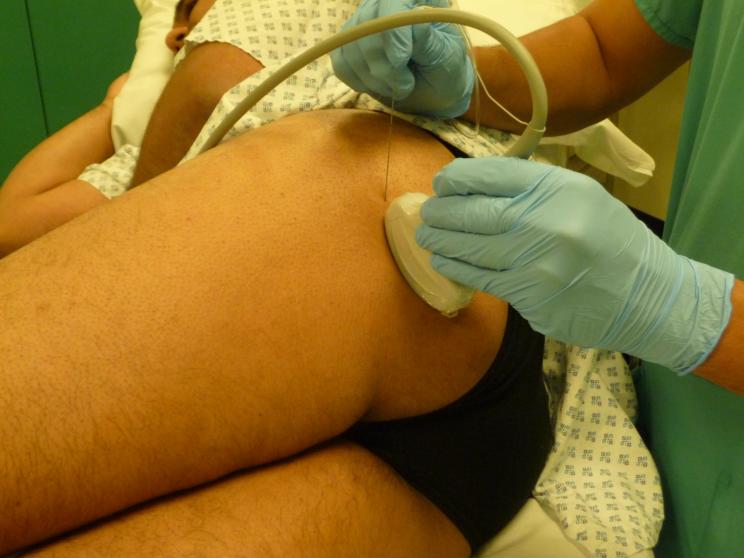
Just above the
gluteal crease:
At this level the nerve passes between the greater trochanter laterally and
ischial tuberosity medially. It lies underneath the gluteus maximus
muscle and it lies between the gluteus and a smaller quadratus femoris muscle.
A curvilinear ultrasound probe (2-6 MHz) is used to visualise the nerve at
this level. Bony shadows produced by the greater trochanter will be seen
laterally and ischial tuberosity will be seen on medial side. You will see a
fascial covering bony shadows going from one bony prominence to other like a
hammock. You will see the sciatic nerve resting on this hammock between
gluteus maximus and quadratus femoris muscle.
This picture shows how the patient is positioned and how the ultrasound probe
is placed.
Following video shows how to locate the nerve:
This video shows the block being done.
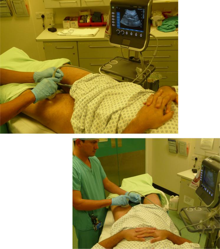
Just below the gluteal crease:
At this level the nerve lies sandwitched between two muscle layers. One
layer is formed by the semimembranosus and semitendinosus muscles medially and
biceps femoris muscle lying on the lateral side. This layer is
superficial when the ultrasound probe is placed posteriorly. The deeper
layer is formed by the adductor magnus muscle.
When the probe is positioned as shown in the picture,
Following will be seen:

Follwoing video from RA-UK show subgluteal sciatic block being done:
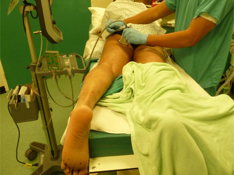
Anterior approach to sciatic nerve:
In this approach with patient supine, a curvilinear probe is placed as shown
in the picture at just above the midthigh level.
You will see bony shadow of the femur with femoral vessels seen superficially.
Three muscle layers will be seen as seen in the following animations:
First animation shows where the probe is and which muscle groups I am talikng
about:
Second animation again shows the same point but also shows the position of the
probe.
Follwoing video shows an anterior sciatic nerve block:


