Popliteal block
The popliteal block is used to provide analgesia for
operations on the ankle.
The advantage of this block over the proximal
approaches to the sciatic nerve are that the hamstrings are spared.
The block can
be used for forefoot operations but an ankle block is preferred technique as it
does not cause foot drop.
Anatomy:
 Click
on image for larger image
Click
on image for larger image
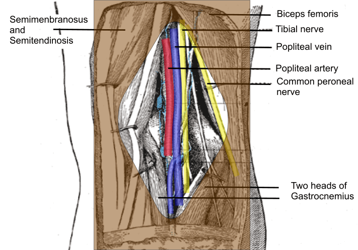
1. The popliteal fossa
is a diamond shaped region at the back of the knee. The superomedial boundary
is made up of semimembranosus and semitendinosis muscle while the superolateral
boundary is made by biceps femoris muscle.
The two heads of gastrocnemius form
the inferolateral and medial boundary.
The Neurovascular bundle lies in the
fossa covered by skin and subcutaneous tissue.
2. The popliteal vein and
artery are deeper (away from skin) compared to the nerves when looked at from
behind the knee.
3. The sciatic nerve divides into its two terminal (Tibial
and Common peroneal) branches 5 to 12 cm above the popliteal crease.
4.
The nerves are always superficial (closer to the skin) than the artery and vein.
Compared to the common peroneal nerve the Tibial nerve lies closer to the artery. It is superficial and lateral to the
artery.
The common peroneal nerve lies lateral to the tibial nerve on the inner
side of biceps femoris muscle.
5. The tibial nerve is always larger than the
common peroneal nerve.
6. In most patients (Depending on the circumference
of the thigh) the nerves are within 2-4 cm of the skin.
7. The medial side
of the leg and foot is innervated by the saphenous nerve so that this nerve will
need to be blocked separately.
Indications:
1. Surgery on the ankle (e.g. ankle fusion or
replacement) and foot.
Although for forefoot operations an ankle block is more
appropriate.
A proximal sciatic block will affect the hamstring muscles, which will
be spared with popliteal block.
A popliteal block will always cause foot drop
though.
Contraindications:
1. Patient refusal
2. Coagulopathy
3. Infection at the site of the block.
4. If patient needs to be mobilized
immediately after operation then foot drop may be a problem.
Key points:
1. Start scanning just above the popliteal crease and follow the nerves cephalad.
2. The thigh is not a perfect cylinder and nerves do not travel in a straight
line in it.
In order to get the best picture of nerve the probe will have to be
angled cephalad and caudal and rotated clockwise and anticlockwise.
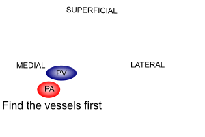
3. Look
for the blood vessels first. The nerves are always superficial and lateral to
them.
Necessary Equipment:
1. Ultrasound machine with high frequency
probe, probe cover and ultrasound gel
2. Insulated stimulating needle (I use 100 mm stimulating needle)+/- nerve
stimulator
3. Local anaesthetic: I use 20 ml of 0.5% levobupivacaine if block is done
for anaesthesia. For postoperative analgesia 0.25% levobupivacaine is also
sufficient
4. 2% chlorhexidine to clean the skin
Locating the nerve:
The ultrasound probe is
placed transversely across the popliteal fossa, just above the crease.
Anticipated depth of the nerves is between 2-4 cm and the ultrasound depth is
set accordingly.
 |
Do not try to see the nerves first. |
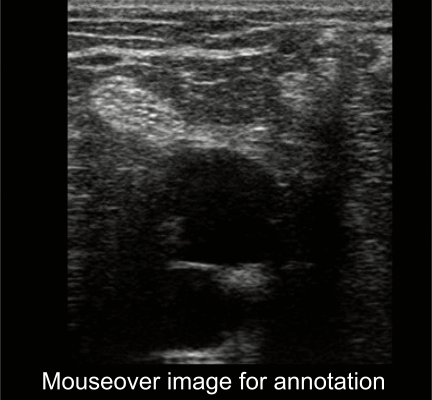 |
The artery (Hypo echoic round structure) is at
the bottom. |
Mouseover image for annotation.
You can see the nerves do see-saw with foot movement in the video below.
Performing the block:
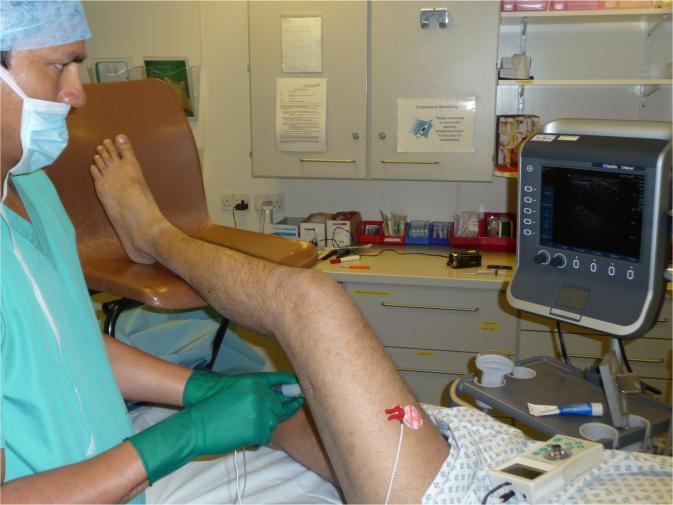
If the patient is awake I do this block with patient in lateral
position.
The leg to be blocked is uppermost and the ultrasound machine is
placed such that the screen is easily visualized.
In an anaesthetized patient, the block is done with patient supine, breathing
spontaneously on LMA.
The patients
leg rests on a chair. Note the position of patient, anaesthetist and
machine in the picture.
I stimulate both the nerves individually under ultrasound
guidance.
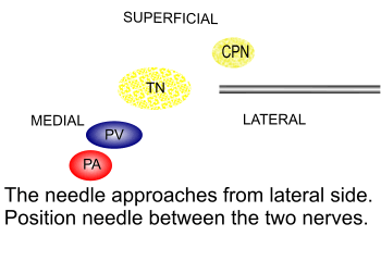
I approach the nerves in plane.
I approach the nerves at a point
where they are near each other.
 |
Use 20 ml of local anaesthetic. |
Nerve stimulation
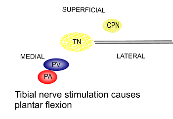 |
Tibial nerve stimulation causes plantar flexion |
The following video demonstrates an ultrasound guided popliteal nerve block.
This
video is courtesy of 'Nerve Imaging Group'.


